

How to assess breathing changes in the non-deteriorating patient as part of a comprehensive respiratory assessment
Abstract
A respiratory assessment seeks to identify changes to respiratory function. Recognising signs of deterioration and escalating a patient’s care can prevent emergencies and reduce further distress for the patient. This article, the fourth in a series on assessment and interpretation for advanced clinical practitioners, outlines a systematic approach to assessing breathing changes in the non-deteriorating patient.
Citation: Cockerill E (2024) How to carry out a respiratory assessment in advanced practice. Nursing Times [online]; 120: 5.
Author: Elena Cockerill is lecturer in physiotherapy, University of Hull.
- This article has been double-blind peer reviewed
- Scroll down to read the article or download a print-friendly PDF here (if the PDF fails to fully download please try again using a different browser)
- Click here to see other articles in this series
Introduction
A respiratory assessment is a systematic analysis of breathing to identify changes to respiratory function. This requires an understanding of the anatomy and physiology of the respiratory system (Cedar, 2018; Hartley, 2018). Fig 1a illustrates the anatomy of the lungs and Fig 1b shows the normal movement of the chest wall when a person is breathing.

Timely respiratory assessment allows for appropriate interventions to mitigate breathing difficulties and prevent further deterioration (Day, 2022). There are three key stages:
- Checking the medical notes;
- Patient questioning (subjective);
- Clinical examination (objective) (Curr and Fordham-Clarke, 2022).
Check the medical notes
Look for a history of the presenting condition and check the details, along with past medical history, as not all causes of respiratory distress arise in the respiratory tract. Heart failure, neuromuscular disorders, toxic ingestion and central nervous system disorders may all manifest with respiratory signs and symptoms (Stephany, 2022); as an example of this, neuromuscular disorders such as motor neurone disease can cause a weak cough (Miller et al, 2017). Obesity can cause mechanical compression of the respiratory system and restrictive pulmonary damage (Mafort et al, 2016), while cardiac conditions can cause breathlessness.
Post-operative patients are at risk of respiratory complications. Changes to the respiratory system that occur immediately on induction of general anaesthesia include decreased respiratory drive, altered muscle function, reduced lung volumes and atelectasis (lung collapse); these increase the risk of post-operative pneumonia (Miskovic and Lumb, 2017). Abdominal or chest surgery can cause pain when coughing and inhibit taking a deep breath, which reduces tidal volumes and further increases the risk of atelectasis and chest infection (Hough, 2017).
Consider the individual’s social and family history for relevant risk factors. Smoking or vaping history is also clinically relevant as both increase the risk of certain lung pathologies even if the person no longer smokes or vapes (Xie et al, 2020; O’Keeffe et al, 2018).
Check whether the patient has been admitted to hospital before with the same presentation. This will help to give an overview of the condition and help you to ascertain where on the disease trajectory the patient could be.
Consult the patient’s notes for their wishes around resuscitation and escalation of care should they deteriorate. This will help to make sure the care they receive is appropriate.
Patient questioning
Use patient questioning to ascertain the main problem or symptom. Open-ended questions allow patients to communicate their concerns in their own words but, if breathlessness makes it difficult for a person to communicate, closed questions can be useful for key aspects of the assessment. When communication is a problem, a collateral history from family members or carers can be used to advocate a person’s wishes or needs. If a patient is deteriorating, you may need to progress straight to the patient examination.
Table 1 shows the most common respiratory symptoms along with questions to ask to establish their presence and how long they have manifested. Consider the onset, timing, exacerbating or relieving factors and severity of the symptoms experienced (Curr and Fordham-Clarke, 2022). Some long-term conditions may cause daily symptoms; establishing a patient’s baseline can help you distinguish between changes that are normal for that person and those that may indicate decline or a change in condition.

Interpretation
Use the answers to these questions to inform the patient examination and treatment; for example, if a patient is reporting a productive cough with thick sputum, it could be a sign of a chest infection.
Questioning a patient about chest pain can help you recognise and escalate serious pathology; for example, pain on inspiration may indicate pulmonary embolus, while central chest and radiating pain could indicate a serious cardiac problem needing immediate escalation (Resuscitation Council UK (RCUK), nd). ‘Red-flag’ symptoms for a concerning underlying pathology, such as lung cancer, can include weight loss, persistent cough and haemoptysis (coughing up blood) (Prado et al, 2023).
Long-term respiratory conditions and breathlessness can present differently in different people, with individuals experiencing varying intensity of symptoms even for the same respiratory problem. Having a ‘toolbox’ of techniques and treatments can help you cater for different patient needs.
Allow patients to express their opinions and concerns, and try to establish what their expectations are; this requires going beyond asking them about their symptoms to asking them about their problems and needs (National Institute for Health and Care Excellence (NICE), 2019). Questions could include:
- What is troubling you most at the moment?
- What would you like me to understand about your condition?
- What are your beliefs about your condition?
Working collaboratively with the patient to create a shared understanding of the condition can serve as the basis for ongoing assessment and treatment. Some patients will have lived with a condition for many years, which may have given them some expertise in self-managing it.
Clinical examination
Prepare the patient as follows:
- Explain what the examination will entail and answer any questions;
- Gain the patient’s informed consent, particularly as you may need to loosen or remove their clothing while examining their chest. If a person lacks capacity to consent, decide whether you need to proceed in the patient’s best interests to ensure they receive the care they need;
- Ensure the environment is private and maintain the patient’s dignity throughout by making sure that areas of the body that are not being assessed are covered. A quiet and private environment is not only essential for the patient, but also helps the practitioner to gather and interpret information during the assessment (Sarkar et al, 2015);
- If appropriate and possible, make sure the patient is upright – this increases lung volumes and will allow you to access the anterior and posterior chest wall for auscultation (Hough, 2017). Consider whether the patient needs help and/or support to move into, and maintain, a sitting position;
- Ensure effective communication throughout the examination. Respiratory symptoms (for example, cough, breathlessness, wheeze and fatigue) can exacerbate during the assessment so, where necessary, offer the patient breaks or a change of position to maintain their comfort.
Initial assessment
First, ascertain the severity of the respiratory problem and whether immediate treatment is needed. For a patient who is acutely deteriorating, use the airway, breathing, circulation, disability, exposure (ABCDE) systematic approach (RCUK, nd). Once personal and patient safety is established, the first stage is to complete an airway assessment to identify whether the patient has an airway obstruction and needs emergency care (Hill, 2021).
Although the ABCDE approach is used in resuscitation emergencies, it can also be adapted for routine care (Cathala and Moorely, 2020). The breathing element of this approach gives a logical structure (‘look, listen and feel’) to comprehensively assess breathing changes in a patient who is stable. This includes observing and listening to breathing, followed by palpation of the chest wall (RCUK, nd). A stepwise approach to assessing breathing changes in a patient who is not deteriorating is described below.
Face (look)
Inspect the face for signs of respiratory distress and pain, such as grimacing, gasping or nasal flaring. Patients may also pucker or purse their lips to help reduce the effort needed to breathe.
Check the skin at their lips, or around their mouth, for any blueness or discolouration that indicates cyanosis; this can also be evident on the hands and feet. In a patient who is acutely unwell, cyanosis is an indicator of low oxygen levels and should be treated with high-flow oxygen and escalated immediately (RCUK, nd).
Chest movement (look)
Signs of respiratory distress can include increased respiratory rate and changes to chest movement, breathing pattern and rhythm. Such changes can indicate increased stress on the respiratory system and are an early warning sign for deterioration (Hartley, 2018). Observe the following:
- Chest symmetry – are both sides of the chest expanding equally?
- Chest and abdominal movement – is the chest rising in synchrony with the abdomen?
- Depth of chest movement – is the chest expanding all the way to the bottom of the lungs?
- Accessory muscle use – is the patient using muscles in the neck to lift their shoulder girdle and ribs?
- Breathing rhythm – is the breathing pattern regular?
- Report any signs of abnormal breathing to senior clinicians for immediate investigation due to the risk of deterioration (Wheatley, 2018a).
Patient observations (look)
Accurate and timely observations are an essential part of respiratory assessment and highlight those patients who need respiratory management. The patient’s oxygen saturation level and respiratory rate are particularly relevant to respiratory function (Hartley, 2018), and any deviation from normal should be reported and escalated.
Oxygen saturation
Pulse oximetry indicates the percentage of oxygen that is carried by the red blood cells in the bloodstream, which is a measure of blood oxygen saturation (SpO2). SpO2 can be defined as the ratio of oxyhaemoglobin to the total concentration of haemoglobin present in the blood – 96-99% is the normal level for adults at sea level (Main and Denehy, 2016).
Target ranges for SpO2 include:
- 94-98% for a patient who is acutely ill;
- 88-92% for a patient with chronic obstructive pulmonary disease (COPD) or a patient at risk of retaining carbon dioxide (CO2) (O’Driscoll et al, 2017).
SpO2 outside of the patient’s target range requires escalation for a review of oxygen requirements. Oxygen should be prescribed initially to achieve a normal or near-normal oxygen saturation in most patients who are acutely ill with a normal or low arterial CO2. The concentration of oxygen needed depends on the condition that is being treated (NICE, nd).
When assessing a patient, first establish:
- Whether an oxygen device is being used;
- The oxygen needs of the patient.
For example, if the SpO2 is in the normal range but the patient is on a high amount of oxygen to maintain oxygenation, this may demonstrate a respiratory or cardiac problem. It is crucial to understand the different oxygen devices, and how much oxygen and flow is being delivered to the patient, while ensuring accurate documentation in the patient observation chart. O’Driscoll et al’s (2017) British Thoracic Society Guideline for Oxygen Use in Adults in Healthcare and Emergency Settings advises on how to manage and monitor the patient to keep within the target SpO2.
Respiratory rate (RR)
RR is a measure of ventilation, and it is important to know how to take an accurate RR (Wheatley, 2018b). Normal RR values are 12-16 breaths per minute (NICE, 2020). RR will increase if there is inadequate gaseous exchange; >30 breaths per minute could be a sign of respiratory failure (O’Driscoll et al, 2017). Conversely, a low RR could be caused by oversedation or a neurological problem (Hough, 2017). Establish baseline observations and trends; chronic lung conditions can cause a higher resting RR to maintain gaseous exchange.
Other vital signs
Heart rate and blood pressure are important observations, as cardiac problems can affect respiratory function.
Patient temperature also informs the assessment as pyrexia (fever) could indicate respiratory infection, requiring further investigation and management.
A drop in consciousness can negatively affect respiratory drive and function, and is an important consideration for escalating patient care.
Complete the National Early Warning Score (NEWS 2) chart – a system for scoring physiological measurements to assess illness severity and risk of deterioration – to help manage, in a timely fashion, a patient who is deteriorating (NICE, 2020).
Position of the patient
Observe the position of the patient because lung volumes reduce in patients who are in supine or semi-recumbent positions, compared with when they are standing or sitting (Katz et al, 2018). Consequently, patients who are breathless may find that lying flat makes their symptoms worse (Cross et al, 2020). Patients may lean forwards or ‘fix’ with their arms to catch their breath (similar to fixing the arms on the thighs after running). This facilitates the use of accessory muscles that help to pull the ribs further outwards, thereby increasing lung volumes, but prolonged use of these muscles can cause fatigue.
Cough and sputum
The type of cough a patient has may also help to ascertain the respiratory problem. For example, a productive cough, whereby sputum is expectorated, can be caused by a chest infection; conversely, a dry cough (as in asthma or with Covid-19) is more likely to be caused by irritation or a narrowing of the airways.
If the patient is expectorating sputum, assess its colour, amount and viscosity (Fig 2). If a patient with a productive cough is struggling to expectorate, this could indicate an ineffective cough or very thick sputum. Medications, such as mucolytics, nebulisers, and humidification of oxygen, can all help to thin sputum so the patient can expectorate more easily. Patient hydration will also reduce the viscosity of any secretions.

Voice (listen)
Listening to the patient’s speech can give vital information about respiratory function. The ability to speak in full sentences is a good indicator of breathing status; taking breaths mid-sentence indicates that a patient is working harder to breathe.
Patients experiencing a large increase in sputum, along with difficulties coughing and clearing, may have sputum vibrating in their upper airways when they breathe or speak, causing a rattling or rasping sound.
Lungs (listen)
An essential next step is auscultation of the chest because it will help you to identify what the respiratory problem could be (Proctor and Rickards, 2020). Auscultation is a structured process of using a stethoscope on the skin to assess airflow throughout the lung fields (Sarkar et al, 2015). Fig 3 highlights the auscultation points to cover with the stethoscope.

The advised technique to use when performing auscultation is to listen to all of the lung fields on full inspiration followed by expiration, and to finish by comparing the left and right lung fields with each other (Proctor and Rickards, 2020). Ideally, make sure the patient is in a sitting position and, when you are listening posteriorly, move their arms forward as this will move their scapulae (shoulder blades) out of the way.
Palpation (feel)
Palpation evaluates chest expansion, depth of breathing and symmetry. Feeling the movement of the chest indicates how well the lungs expand and recoil.
Lung expansion and symmetry
Various pathologies can affect chest expansion, as the following examples show:
- COPD causes reduced recoil and hyperinflation of the chest;
- Kyphoscoliosis (abnormal curvature of the spine in the lateral and forward direction) and chest wall deformities cause restriction to the chest wall – this is characterised by reduced lung volumes and breathlessness, making reduced chest expansion on palpation likely (Fuschillo et al, 2015);
- Pneumothorax (air or gas in the cavity between the lungs and chest wall) and lung consolidation (replacement of
air in the small airways with fluid, solid or other material) can cause asymmetry on lung expansion
(Innes and Tiernan, 2018).
Normal chest expansion is typically 2-4cm (Innes et al, 2018). To assess expansion and symmetry, place your hands firmly on the chest wall at the same level and check whether your thumbs move equally apart as the patient breathes in, feeling for asymmetric movement (Shellenberger et al, 2017). Perform this sequentially and systematically anteriorly and posteriorly, working from the apex down to the bases of the lungs (Fig 4).
![]()
Chest wall palpation
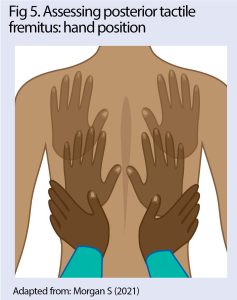
Changes in sound vibrations in the chest wall (tactile fremitus) can signify abnormalities of the lung tissue, such as collapse or consolidation (Rao et al, 2019). Assess tactile fremitus by placing the flat palm of the hand on the chest, while asking the patient to say “99”. Perform this sequentially anteriorly and posteriorly, adopting a systematic approach (Morgan, 2021). Hand positions for posterior assessment are shown in Fig 5.
In a patient that is unable to speak, palpation of the chest wall is a useful tool for assessing for signs of sputum retention, which can be felt as crackles over the affected area or a popping sensation under the skin (Cross et al, 2020). Air trapping between the chest wall and skin, caused by a medical procedure (surgical emphysema) can be felt in the same way.
Chest percussion
Place one finger of your non-dominant hand on the patient’s chest wall, then tap horizontally onto the phalanges of this finger with one or two fingers of your other hand. The sound produced depends on the state of the patient’s underlying lung tissue (Rao et al, 2019). If the lungs or pleura contain sputum or liquid, percussion will produce a dull sound, but pneumothorax produces a hollow note (Rao et al, 2019). Complete this systematically from top to bottom, comparing left with right. When percussing posteriorly, position the patient with their arms forward to move the scapulae our of the way.

Respiratory investigations
Investigations are guided by the findings of the respiratory assessment – for example, an arterial blood gas test can check for developing respiratory failure in patients with high oxygen requirements or deteriorating SpO2 (O’Driscoll et al, 2017), while a chest X-ray can confirm assessment findings and help you diagnose a problem such as pneumothorax, lung consolidation or pleural effusion. Interpretation of chest X-rays is discussed in more detail in a later article in this series.
Conclusion
Respiratory assessment requires a logical structure and consideration of patient comfort and clinical status. Deteriorating patients need assessment to be succinct to escalate the treatment of an airway or breathing problem. For patients who are stable however, a detailed assessment can be made to gain a full understanding of the symptoms and respiratory problem, and allow a collaborative approach to care.
Advanced practitioners
This series is aimed at nurses and midwives working at, or towards, advanced practice. Advanced practitioners are educated at master’s level and are assessed as competent to make autonomous decisions in assessing, diagnosing and treating patients. Advanced assessment and interpretation is based on a medical model, and the role of advanced practitioners is to integrate this into a holistic package of care.
- Professional responsibilities – This procedure should be undertaken only after approved training, supervised practice and competency assessment, and carried out in accordance with local policies and protocols.
Cedar SH (2018) Every breath you take: the process of breathing explained. Nursing Times; 114: 1, 47-50.
Cross J et al (2020) Respiratory Physiotherapy Pocketbook: An On Call Survival Guide. Elsevier.
Curr S, Fordham-Clarke C (2022) Clinical Nursing Skills at a Glance. Wiley Blackwell.
Day K (2022) Essential critical care skills 4: airway assessment and management. Nursing Times; 118: 2, 29-32.
Fuschillo S et al (2015) Pulmonary rehabilitation improves exercise capacity in subjects with kyphoscoliosis and severe respiratory impairment. Respiratory Care; 60: 1, 96-101.
Hartley J (2018) Respiratory rate 2: anatomy and physiology of breathing. Nursing Times; 114: 6, 43-44.
Hill K (2021) Essential critical care skills 2: assessing the patient. Nursing Times; 117: 12, 35-38.
Hough A (2017) Hough’s Cardiorespiratory Care: An Evidence-based, Problem-solving Approach. Elsevier.
Innes JA et al (eds) (2018) The respiratory system. In: Innes JA et al (eds) Macleod’s Clinical Examination. Elsevier.
Katz S et al (2018) The effect of body position on pulmonary function: a systematic review. BMC Pulmonary Medicine; 18: 159.
Mafort TT et al (2016) Obesity: systemic and pulmonary complications, biochemical abnormalities, and impairment of lung function. Multidisciplinary Respiratory Medicine; 11: 28.
Main E, Denehy L (2016) Cardiorespiratory Physiotherapy: Adults and Paediatrics. Elsevier.
Miller S et al (2017) Assessment of airway defenses in the neurologically impaired patient. Medsurg Nursing; 26: 2; 113-118.
Miskovic A, Lumb AB (2017) Postoperative pulmonary complications. British Journal of Anaesthesia; 118: 3, 317-334.
Morgan S (2022) Respiratory assessment: undertaking a physical examination of the chest in adults. Nursing Standard; 37: 3, 75-82.
National Institute for Health and Care Excellence (nd) Oxygen. bnf.nice.org.uk (accessed 2 April 2024).
National Institute for Health and Care Excellence (2020) National Early Warning Score Systems that Alert to Deteriorating Adult Patients in Hospital. NICE.
National Institute for Health and Care Excellence (2019) Chronic obstructive pulmonary disease in over 16s: diagnosis and management. nice.org.uk, (accessed 2 April 2024).
O’Driscoll BR et al (2017) BTS guideline for oxygen use in adults in healthcare and emergency settings. Thorax; 72: ii1-ii90.
O’Keeffe LM et al (2018) Smoking as a risk factor for lung cancer in women and men: a systematic review and meta-analysis. BMJ Open; 8: e021611.
Prado MG et al (2023) Symptoms and signs of lung cancer prior to diagnosis: case–control study using electronic health records from ambulatory care within a large US-based tertiary care centre. BMJ Open; 13: e068832.
Proctor J, Rickards E (2020) How to perform chest auscultation and interpret the findings. Nursing Times; 116: 1, 23-26.
Rao A et al (2019) Acoustic methods for pulmonary diagnosis. IEEE Reviews in Biomedical Engineering; 12: 221-239.
Resuscitation Council UK (nd) The ABCDE approach. resus.org.uk (accessed 7 March 2024).
Sarkar M et al (2015) Auscultation of the respiratory system. Annals of Thoracic Medicine; 10: 3, 158-168.
Shellenberger RA et al (2017) Diagnostic value of the physical examination in patients with dyspnea. Cleveland Clinical Journal of Medicine; 84: 12, 943-950.
Stephany A (2022) Respiratory distress. In: Kliegman RM et al (eds) Nelson Pediatric Symptom-based Diagnosis: Common Diseases and their Mimics. Elsevier.
Wheatley I (2018a) Respiratory rate 5: using this vital sign to detect deterioration. Nursing Times; 114: 10, 45-46.
Wheatley I (2018b) Respiratory rate 3: how to take an accurate measurement. Nursing Times; 114: 7, 21-22.
Xie W et al (2020) Association of electronic cigarette use with incident respiratory conditions among US adults from 2013 to 2018. JAMA Network Open; 3: 11, e2020816.
Help Nursing Times improve
Help us better understand how you use our clinical articles, what you think about them and how you would improve them. Please complete our short survey.
link